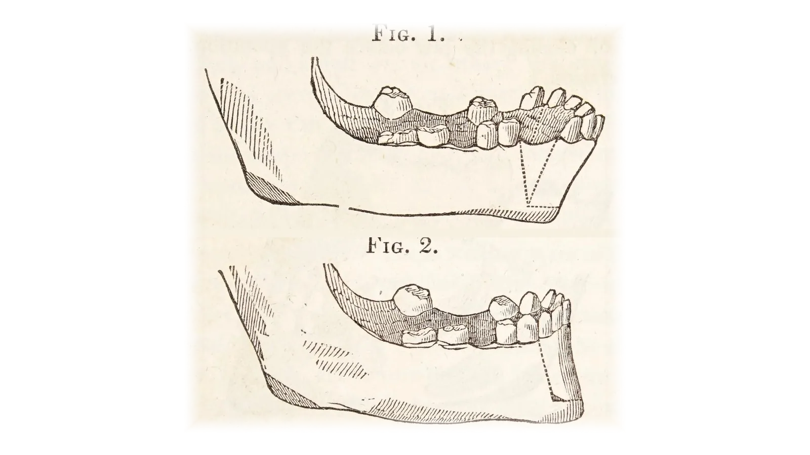
Hullihen, 1849: the first published dentoalveolar osteotomy.
The evolution of orthognathic surgery
From plaster and paper to virtual planning: this is how we got here.
Where it began
Orthognathic surgery began with osteotomies, precise cuts in the bone, so that the teeth could bite properly. Over time, the goal grew: repositioning the upper jaw (maxilla) and the lower jaw (mandible) not only for the bite, but also to improve the face. That planning was done with plaster models and tracings on paper over X-rays, measuring by hand and taking those measurements to the operating room. It was a great advance for its time, and everything that came after was built on that foundation.
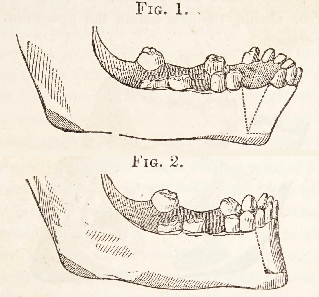
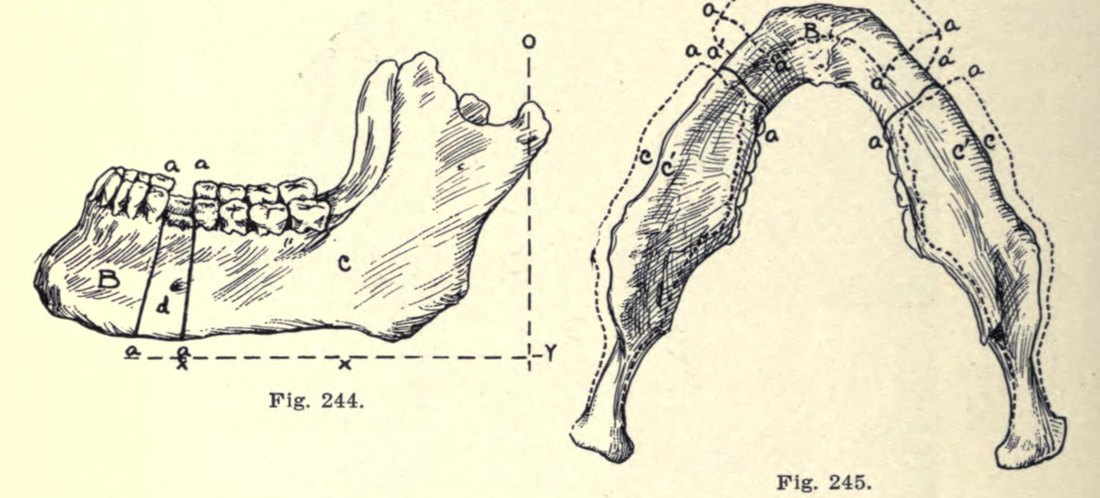
From wires to plates
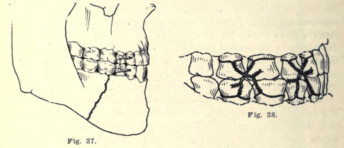
For a long time, after surgery, the jaws were held together with wires and the mouth stayed closed for weeks. The arrival of plates and screws changed that: the bones are held firmly in their new position, you can talk and open your mouth from the first day, recovery is faster and there's more confidence in the results. Later, photographs were added to the study of each case, to look at the face in more detail.
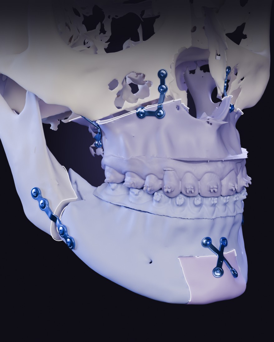
Virtual planning
Virtual planning opened up new possibilities, and today it's the standard. With your 3D scans and a scan of your teeth, a digital twin of your face is built: bones, bite, joints and airway. On that twin, the surgery is first performed virtually, reviewing each option with its benefits and its limits. Then, guides printed just for you make it possible to reproduce that plan with precision in the operating room. Nothing is improvised: it's planned. That means fewer surprises and a peace of mind that didn't exist before.
Minimally invasive surgery
Minimally invasive surgery brought new ways of performing the techniques and of caring for recovery. In general, the work is done from inside the mouth, with no visible scars on the face, and instruments like the piezoelectric device, which cuts bone using ultrasound, better protect the sensitive structures around it, such as nerves. All of this translates into something concrete: a calmer recovery and, usually, going home the next day.
Sleep apnea and 3D diagnosis
Orthognathic surgery also found its place in obstructive sleep apnea. When the growth and arrangement of the structure of the face (the jaws, the muscles and, above all, the tongue) leave little room to breathe, moving the jaws forward widens the upper airway, and because the structure changes, that change is permanent. At the same time, 3D diagnosis makes it possible to see beyond a flat X-ray and to measure in volume. Together, they have expanded what surgery can do and improved its results.
Orthofacial: the whole face
Orthofacial surgery brought a more comprehensive view, in two ways. In how it's thought out: from the bite to the whole face. How you bite, how you breathe, how you look and how you feel are all one thing, and the plan considers the harmony between nose, lips and chin, the airway, the joints and stability over time. And in what's done: jaw surgery is joined by other procedures of the face, such as rhinoplasty, facelift and other facial techniques, which are usually assessed, planned and performed together. Everything is chosen case by case to achieve more harmonious results, and the work is done as a team when the case calls for it. It's a field that keeps evolving: today we can do things that used to be impossible; everything else we'll go over at your consultation.
All of this serves one purpose: to understand your case clearly. The first step is an evaluation.
Frequently asked questions
Is orthognathic surgery still major surgery?
Yes: it's performed in the operating room, under general anesthesia and with a full team. What has changed is how it's done and how it's experienced. Today it's planned virtually before surgery, the work is generally done from inside the mouth with minimally invasive techniques, and recovery is faster and calmer than before. Every detail of your case is discussed at your consultation.
Are the jaws still wired shut after surgery?
In most cases, no. Today the bones are held in place with small plates and screws, and you can talk and open your mouth from the first day. Sometimes soft elastics are used for a while to guide the bite; your team will tell you how and when.
What does "orthofacial" mean?
It's a more comprehensive view of surgery: planning the whole face, not just the bite. How you bite, how you breathe, how you look and how you feel are considered together, because in your face they work together. It also widens what's done: alongside jaw surgery, other procedures of the face, such as rhinoplasty or a facelift, which are usually assessed, planned and performed together. Techniques are chosen case by case to achieve more harmonious results.
What is virtual planning?
It's performing your surgery first on the computer, on a digital twin of your face made from your own 3D scans. There, the options are reviewed and the best one for your case is chosen. Today it's the standard, and guides printed just for you help reproduce that plan with precision in the operating room.
Can orthognathic surgery help with sleep apnea?
Yes, when it's properly indicated. If the growth and arrangement of the structure of the face leave little room to breathe, moving the jaws forward widens the upper airway permanently, and it's the surgery with the greatest impact on obstructive sleep apnea. Sleep apnea is diagnosed with a sleep study (polysomnography) and other tests, and it's handled as a team when the case calls for it.
Does the surgery leave scars on the face?
In general, the work is done from inside the mouth, so there are no visible scars on the face. If your case needed a different approach, we would discuss it ahead of time at your consultation, so you know exactly what to expect.
Do you know someone who should read this?
Share it on WhatsApp or Instagram. It's information to help understand, not a diagnosis.